

Hi Ahmed,
I think you are set to go
- First our patient
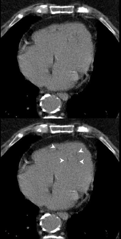
- subtle linear fat accumulation in the septum in this patient with CAD
- Second
- show examples of fat in the heart
There are a lot of cases – Do not feel the need to complete in 3 minutes
Clinical summary of a 65 year old female with longstanding history of SLE, Lupus Sjogren’s and Raynaud’s presented with 2 weeks of dyspnea and elevated troponins suggestive of a STEMI. Cardiac cath showed 2 vessel disease and she was referred for CABG. At surgery there were adhesions and the surgeon was unable to identify the coronaries as a result of the fibrosis. She was closed without surgery. She subsequently had a diagnostic MRI and endomyocardial biopsy which showed chloroquine related cardiomyopathy
Ashley Davidoff MD
Normal and Normal Variants
Epicardial and Pericardial Fat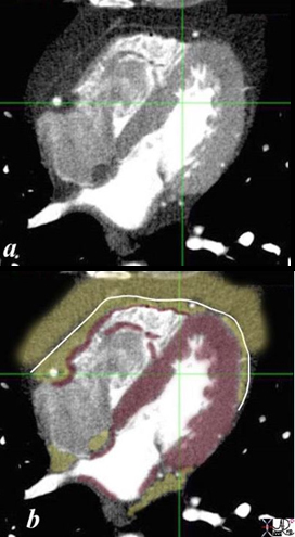
THE PERICARDIUM , EPICARDIAL FAT AND PERICARDIAL FAT CT scan through the 4 chambers shows a normal pericardium (white line) surrounded by an inner lining of epicardial fat (yellow in contact with the red myocardium ) and an outer layer of pericardial fat) . Note that the coronary arteries (white dots run in the epicardial fat. Ashley Davidoff MD heart anatomy P 08
Large Amount of Epicardial and Pericardial Fat associated with
-
- increased cardiovascular risk, especially for coronary artery disease. – Metabolic Syndrome
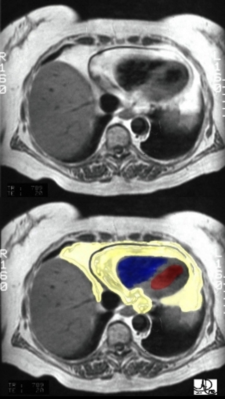
An axial T1 weighted MRI shows a large amount of fat (yellow overlay) around the heart. The epicardial fat is the inner layer and is intimately related to the heart and the pericardial fat is the outer layer. The normal pericardium is seen as a black line between the two layers of fat. Although the pericardium looks like a single layer, it actually consists of two structures – the epicardial serous component and the pericaricardial fibrous component. In pericardial effusion the two layers are separated.
The amount of fat is more than expected and this amount is often associated with Syndrome X.
Courtesy of Ashley Davidoff M.D. 32141.8 code normal heart epicardial fat pericardium anatomy cardiac imaging radiology MRI
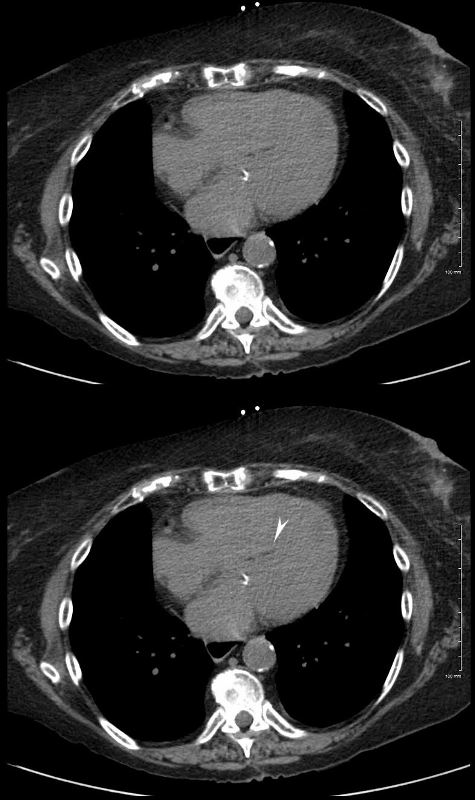
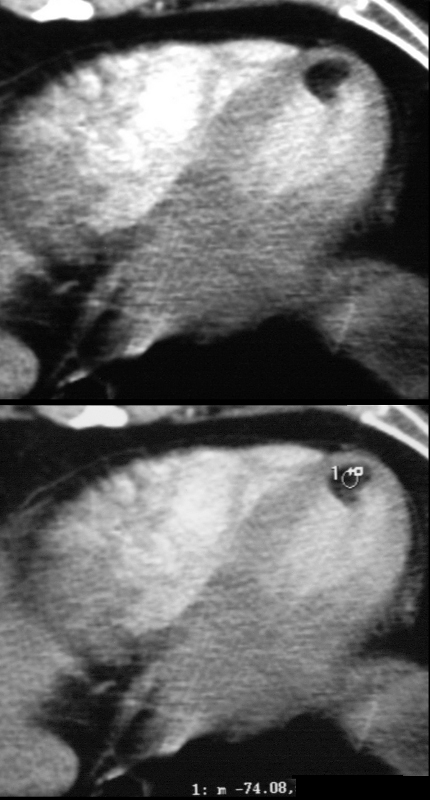
Epicardial Fat with Mild Pressure Effect on the RV
CT scan through the RV and LV shows epicardial fatty accumulation in the subendocardium of the right ventricle. The normal pericardium, (white arrowhead) and normal epicardial fat (yellow arrowhead) are shown in the lower images.
Ashley Davidoff MD
130641L
Degenerative and Age Related Changes in the RV
Axial, non contrast CT scan of a 100 year old female showing physiological accumulations of fat in the right ventricle (RV) and likely in the papillary muscles of the left ventricle (LV)
Ashley Davidoff MD
In the RV Wall
Ashley Davidoff MD
130639L
Atrial Septal Lipoma and Large Amount of Pericardial Fat Patient on Steroids
Axial CT scan through the heart is from a 71year old female who is on steroid therapy. A large amount of fat is noted in the epicardium (yellow arrowhead) and also in the interatrial septum resulting in an atrial lipoma.
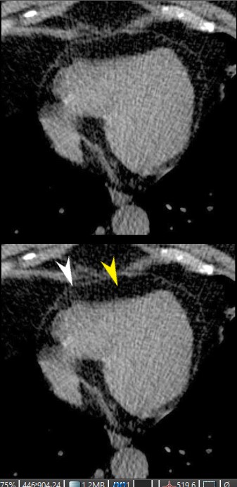
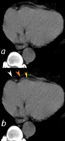
In CAD and prior Myocardial Infarction
56 year old male with history of coronary artery disease. Axial CT through the heart shows apical curvilinear fat (yellow arrowheads, ( a and b) associated with apical myocardial dystrophic calcification (green arrowheads c and d) both indicating prior apical MI. In addition there mitral annular calcification (red arrowhead, b) and multifocal fatty deposits in the RV (white arrowheads, a and b) usually depicting age related degenerative changes,
Ashley Davidoff MD
Unusual cases
Pericardial Lipoma
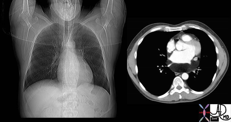
LIPOMA OF THE PERICARDIUM
CXR shows widening of the left main bronchus and a vague lucency at the apex of the heart, CT shows lipoma posterior to the LA
Lipoma at the SVC RA junction
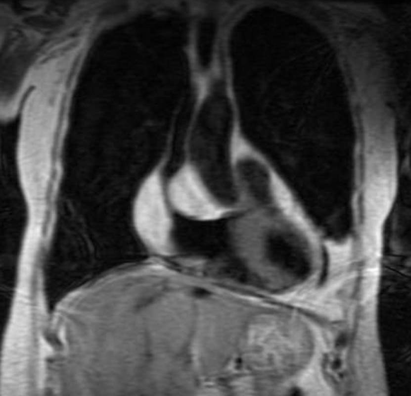
This image of a coronally acquired T1 weighted MRI image of the heart shows a high intensity mass surrounding the SVC and the entrance right atrium (RA) with narrowing of the SVC. There were no symptoms of SVC syndrome in this patient with known COPD. Note that the mass has the intensity of subcutaneous fat. An atrial lipoma is the most likely diagnosis.
Courtesy Jorge Medina
38449c
KEY WORDS
Cardiac, heart, vein, SVC , RA, mass, fat, lipoma, tumor, neoplasm, benign, imaging, radiology, MRI

This series of axial T1 weighted MRI images of the heart show a high intensity mass surrounding the SVC (blue arrowhead) and the entrance to the right atrium (RA). There were no symptoms of SVC syndrome in this patient with known COPD. Note that the mass has the intensity of subcutaneous fat. An atrial lipoma is the most likely diagnosis.
Courtesy Jorge Medina
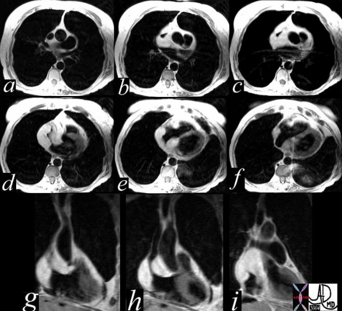
This series of axial and coronal T1 weighted MRI images of the heart show a high intensity mass surrounding the SVC and the entrance to the right atrium. There were no symptoms of SVC syndrome in this patient with known COPD. Note that the mass has the intensity of the subcutaneous fat. An atrial lipoma is the most likely diagnosis.
Courtesy Jorge Medina
38449c
KEY WORDS
Cardiac, heart, vein, SVC , RA, mass, fat, lipoma, tumor, neoplasm, benign, imaging, radiology, MRI
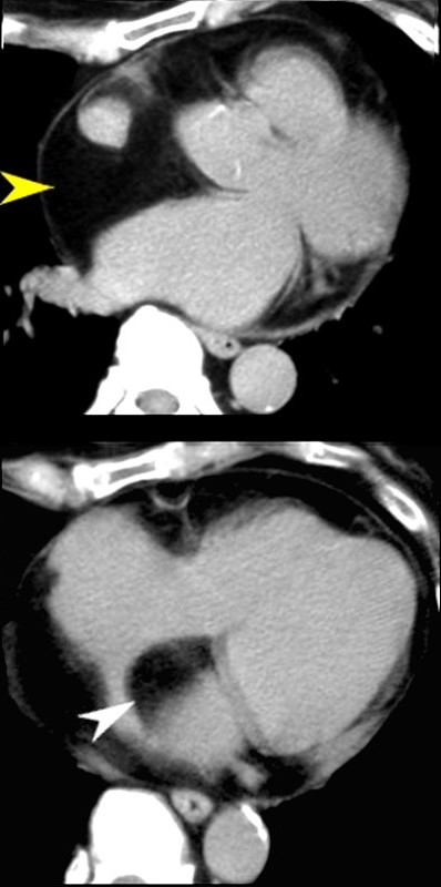
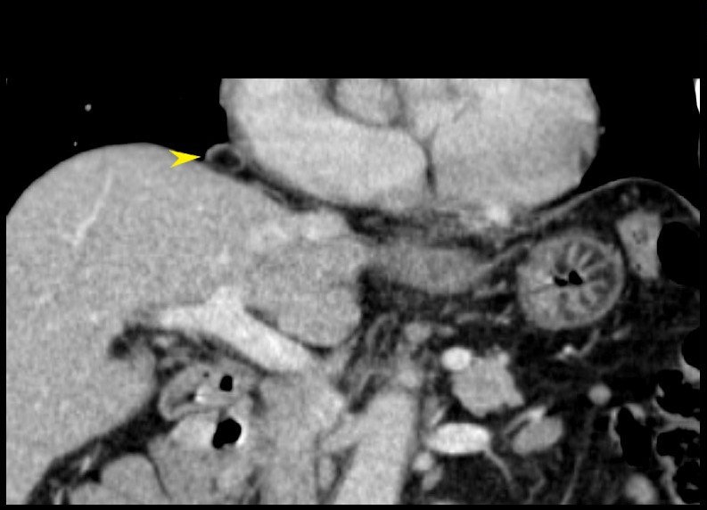
FAT NECROSIS-Target shaped
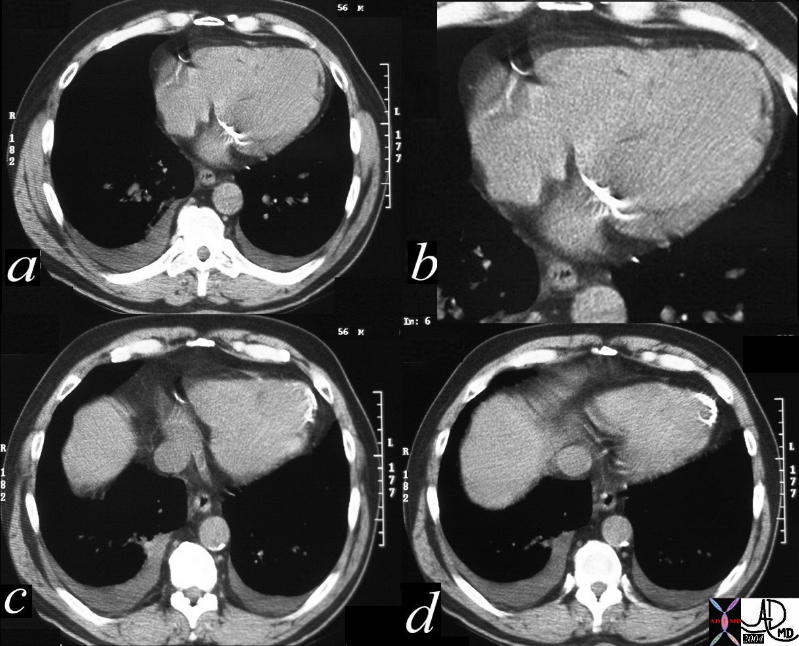
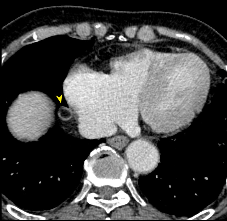
EPICARDIAL FAT NECROSIS
79-year male with asymptomatic finding on axial CT of a focal sclerotic ring surround the epicardial fat on the right side of the heart (yellow arrowhead). This most likely reflects chronic epicardial fat necrosis
Ashley Davidoff MD
79-year male with asymptomatic finding on coronally reformatted CT of a focal sclerotic ring surround the epicardial fat on the right side of the heart (yellow arrowhead). This most likely reflects chronic epicardial fat necrosis
Ashley Davidoff MD
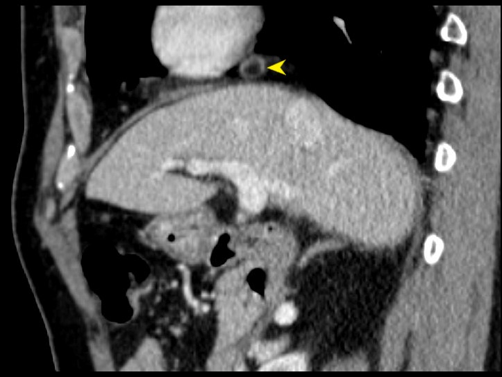
79-year male with asymptomatic finding on sagittally reformatted CT of a focal sclerotic ring surround the epicardial fat on the right side of the heart (yellow arrowhead). This most likely reflects chronic epicardial fat necrosis
Ashley Davidoff MD
Lipoma of the LV
37 year old male with no history of CAD with a fat containing nodule at the LV apex most likely representing a lipoma of the myocardium
Ashley Davidoff MD
Kimura et al Myocardial Fat at Cardiac Imaging: How Can We Differentiate Pathologic from Physiologic Fatty Infiltration? RadioGraphicsVol. 30, No. 6