The Common Vein Copyright 2010
Introduction
Thyroid Technetium Pertechnitate Scan
PET Scan
|
Lymphoma of the Thyroid |
|
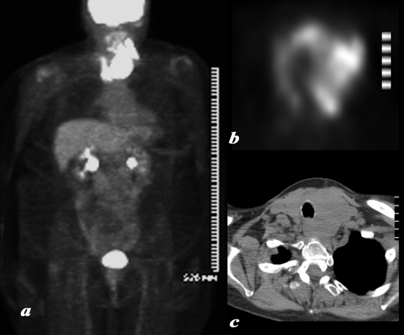
A large and very PET avid left lobe of the thyroid is demonstrated on the body PET scan shown in full body view in (a) and in transverse view of the thyroid in (b). The patient is a 62 year female who had a biopsy proven lymphoma of the thyroid.. The CT shows asymmetric and smooth and homogeneous enlargement of the thyroid. The findings are consistent with the final biopsy proven diagnosis of lymphoma. Courtesy Ashley Davidoff MD Copyright 2010 95855c01.8 |
|
PET following Chemotherapy and Radiation Therapy |
|
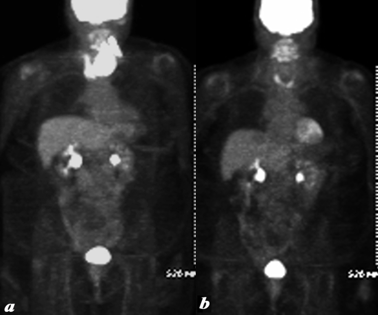
The patient is a 62 year female who had a biopsy proven lymphoma of the thyroid . A large and very PET avid left lobe of the thyroid is demonstrated on the body PET scan shown in full body view in (a) with resolution of the mass and significant and likely remission changes (b) following chemotherapy and radiotherapy. At the base of the neck there is a U shaped pattern corresponding to residual thyroid tissue. The SUV was 5, higher than typically expected which usually ranges between 1and 2 suggesting persistent moderate uptake. This is likely due to radiation thyroiditis. The findings are most consistent with lymphoma in remission following therapy and likely associated XRt thyroiditis. Courtesy Ashley Davidoff MD Copyright 2010 95855c02.8 |
Limitations
—
DOMElement Object
(
    [schemaTypeInfo] =>
    [tagName] => table
    [firstElementChild] => (object value omitted)
    [lastElementChild] => (object value omitted)
    [childElementCount] => 1
    [previousElementSibling] => (object value omitted)
    [nextElementSibling] => (object value omitted)
    [nodeName] => table
    [nodeValue] =>


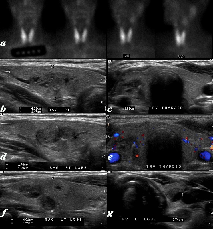
Limitaions in Resolving a 1.8cms Nodule


This is a thyroid scan from a 53 year old female with a clinical history of goiter. She was injected with Iodine -123 . 24 hours later radioiodine uptake was calculated at 31.8% (normal 8-35%). She was also injected with 14.2 mCi of 99mtechnetium pertechnitate.. The scan (a) shows an enlarged gland bilaterally with uniform uptake of the radioisotope in both lobes of the thyroid gland. The uptake in the thyroid gland is normal relative to the uptake in the submandibular and parotid glands. These findings are consistent with a diffuse goiter. The ultrasound on the other hand shows a normal sized gland with multiple nodules. The right lobe (c,d) measures 4.4cms (length) X 1.5cms (A-P) X 1.8cms (TRV) and the left lobe (f,g) measures 4.9cms (length) X 1.6cms (A-P) X 1.1cms (TRV) In the right lobe there is a nodule in the middle of the gland (b,c,d, e) that is relatively hypovascular that measures 1.8cms X 1.1 cms (d,e). Two 7mms nodules were also seen in the left lobe (f,g) Whereas the thyroid nuclear scan was able to accurately depict function, the ultrasound was more accurate in defining the exact size and nodular character of the gland. The diagnosis based on imaging therefore was a non toxic multinodular normal sized gland..
Courtesy Alan Ashare MD and Ashley Davidoff MD Copyright 2010 97513c03.8L


    [nodeType] => 1
    [parentNode] => (object value omitted)
    [childNodes] => (object value omitted)
    [firstChild] => (object value omitted)
    [lastChild] => (object value omitted)
    [previousSibling] => (object value omitted)
    [nextSibling] => (object value omitted)
    [attributes] => (object value omitted)
    [ownerDocument] => (object value omitted)
    [namespaceURI] =>
    [prefix] =>
    [localName] => table
    [baseURI] =>
    [textContent] =>


Limitaions in Resolving a 1.8cms Nodule


This is a thyroid scan from a 53 year old female with a clinical history of goiter. She was injected with Iodine -123 . 24 hours later radioiodine uptake was calculated at 31.8% (normal 8-35%). She was also injected with 14.2 mCi of 99mtechnetium pertechnitate.. The scan (a) shows an enlarged gland bilaterally with uniform uptake of the radioisotope in both lobes of the thyroid gland. The uptake in the thyroid gland is normal relative to the uptake in the submandibular and parotid glands. These findings are consistent with a diffuse goiter. The ultrasound on the other hand shows a normal sized gland with multiple nodules. The right lobe (c,d) measures 4.4cms (length) X 1.5cms (A-P) X 1.8cms (TRV) and the left lobe (f,g) measures 4.9cms (length) X 1.6cms (A-P) X 1.1cms (TRV) In the right lobe there is a nodule in the middle of the gland (b,c,d, e) that is relatively hypovascular that measures 1.8cms X 1.1 cms (d,e). Two 7mms nodules were also seen in the left lobe (f,g) Whereas the thyroid nuclear scan was able to accurately depict function, the ultrasound was more accurate in defining the exact size and nodular character of the gland. The diagnosis based on imaging therefore was a non toxic multinodular normal sized gland..
Courtesy Alan Ashare MD and Ashley Davidoff MD Copyright 2010 97513c03.8L


)
DOMElement Object
(
    [schemaTypeInfo] =>
    [tagName] => td
    [firstElementChild] => (object value omitted)
    [lastElementChild] => (object value omitted)
    [childElementCount] => 2
    [previousElementSibling] =>
    [nextElementSibling] =>
    [nodeName] => td
    [nodeValue] =>
This is a thyroid scan from a 53 year old female with a clinical history of goiter. She was injected with Iodine -123 . 24 hours later radioiodine uptake was calculated at 31.8% (normal 8-35%). She was also injected with 14.2 mCi of 99mtechnetium pertechnitate.. The scan (a) shows an enlarged gland bilaterally with uniform uptake of the radioisotope in both lobes of the thyroid gland. The uptake in the thyroid gland is normal relative to the uptake in the submandibular and parotid glands. These findings are consistent with a diffuse goiter. The ultrasound on the other hand shows a normal sized gland with multiple nodules. The right lobe (c,d) measures 4.4cms (length) X 1.5cms (A-P) X 1.8cms (TRV) and the left lobe (f,g) measures 4.9cms (length) X 1.6cms (A-P) X 1.1cms (TRV) In the right lobe there is a nodule in the middle of the gland (b,c,d, e) that is relatively hypovascular that measures 1.8cms X 1.1 cms (d,e). Two 7mms nodules were also seen in the left lobe (f,g) Whereas the thyroid nuclear scan was able to accurately depict function, the ultrasound was more accurate in defining the exact size and nodular character of the gland. The diagnosis based on imaging therefore was a non toxic multinodular normal sized gland..
Courtesy Alan Ashare MD and Ashley Davidoff MD Copyright 2010 97513c03.8L

    [nodeType] => 1
    [parentNode] => (object value omitted)
    [childNodes] => (object value omitted)
    [firstChild] => (object value omitted)
    [lastChild] => (object value omitted)
    [previousSibling] => (object value omitted)
    [nextSibling] => (object value omitted)
    [attributes] => (object value omitted)
    [ownerDocument] => (object value omitted)
    [namespaceURI] =>
    [prefix] =>
    [localName] => td
    [baseURI] =>
    [textContent] =>
This is a thyroid scan from a 53 year old female with a clinical history of goiter. She was injected with Iodine -123 . 24 hours later radioiodine uptake was calculated at 31.8% (normal 8-35%). She was also injected with 14.2 mCi of 99mtechnetium pertechnitate.. The scan (a) shows an enlarged gland bilaterally with uniform uptake of the radioisotope in both lobes of the thyroid gland. The uptake in the thyroid gland is normal relative to the uptake in the submandibular and parotid glands. These findings are consistent with a diffuse goiter. The ultrasound on the other hand shows a normal sized gland with multiple nodules. The right lobe (c,d) measures 4.4cms (length) X 1.5cms (A-P) X 1.8cms (TRV) and the left lobe (f,g) measures 4.9cms (length) X 1.6cms (A-P) X 1.1cms (TRV) In the right lobe there is a nodule in the middle of the gland (b,c,d, e) that is relatively hypovascular that measures 1.8cms X 1.1 cms (d,e). Two 7mms nodules were also seen in the left lobe (f,g) Whereas the thyroid nuclear scan was able to accurately depict function, the ultrasound was more accurate in defining the exact size and nodular character of the gland. The diagnosis based on imaging therefore was a non toxic multinodular normal sized gland..
Courtesy Alan Ashare MD and Ashley Davidoff MD Copyright 2010 97513c03.8L

)
DOMElement Object
(
    [schemaTypeInfo] =>
    [tagName] => td
    [firstElementChild] => (object value omitted)
    [lastElementChild] => (object value omitted)
    [childElementCount] => 2
    [previousElementSibling] =>
    [nextElementSibling] =>
    [nodeName] => td
    [nodeValue] =>
Limitaions in Resolving a 1.8cms Nodule
    [nodeType] => 1
    [parentNode] => (object value omitted)
    [childNodes] => (object value omitted)
    [firstChild] => (object value omitted)
    [lastChild] => (object value omitted)
    [previousSibling] => (object value omitted)
    [nextSibling] => (object value omitted)
    [attributes] => (object value omitted)
    [ownerDocument] => (object value omitted)
    [namespaceURI] =>
    [prefix] =>
    [localName] => td
    [baseURI] =>
    [textContent] =>
Limitaions in Resolving a 1.8cms Nodule
)
DOMElement Object
(
    [schemaTypeInfo] =>
    [tagName] => table
    [firstElementChild] => (object value omitted)
    [lastElementChild] => (object value omitted)
    [childElementCount] => 1
    [previousElementSibling] => (object value omitted)
    [nextElementSibling] => (object value omitted)
    [nodeName] => table
    [nodeValue] =>


PET following Chemotherapy and Radiation Therapy


The patient is a 62 year female who had a biopsy proven lymphoma of the thyroid . A large and very PET avid left lobe of the thyroid is demonstrated on the body PET scan shown in full body view in (a) with resolution of the mass and significant and likely remission changes (b) following chemotherapy and radiotherapy. At the base of the neck there is a U shaped pattern corresponding to residual thyroid tissue. The SUV was 5, higher than typically expected which usually ranges between 1and 2 suggesting persistent moderate uptake. This is likely due to radiation thyroiditis. The findings are most consistent with lymphoma in remission following therapy and likely associated XRt thyroiditis.
Courtesy Ashley Davidoff MD Copyright 2010 95855c02.8


    [nodeType] => 1
    [parentNode] => (object value omitted)
    [childNodes] => (object value omitted)
    [firstChild] => (object value omitted)
    [lastChild] => (object value omitted)
    [previousSibling] => (object value omitted)
    [nextSibling] => (object value omitted)
    [attributes] => (object value omitted)
    [ownerDocument] => (object value omitted)
    [namespaceURI] =>
    [prefix] =>
    [localName] => table
    [baseURI] =>
    [textContent] =>


PET following Chemotherapy and Radiation Therapy


The patient is a 62 year female who had a biopsy proven lymphoma of the thyroid . A large and very PET avid left lobe of the thyroid is demonstrated on the body PET scan shown in full body view in (a) with resolution of the mass and significant and likely remission changes (b) following chemotherapy and radiotherapy. At the base of the neck there is a U shaped pattern corresponding to residual thyroid tissue. The SUV was 5, higher than typically expected which usually ranges between 1and 2 suggesting persistent moderate uptake. This is likely due to radiation thyroiditis. The findings are most consistent with lymphoma in remission following therapy and likely associated XRt thyroiditis.
Courtesy Ashley Davidoff MD Copyright 2010 95855c02.8


)
DOMElement Object
(
    [schemaTypeInfo] =>
    [tagName] => td
    [firstElementChild] => (object value omitted)
    [lastElementChild] => (object value omitted)
    [childElementCount] => 2
    [previousElementSibling] =>
    [nextElementSibling] =>
    [nodeName] => td
    [nodeValue] =>
The patient is a 62 year female who had a biopsy proven lymphoma of the thyroid . A large and very PET avid left lobe of the thyroid is demonstrated on the body PET scan shown in full body view in (a) with resolution of the mass and significant and likely remission changes (b) following chemotherapy and radiotherapy. At the base of the neck there is a U shaped pattern corresponding to residual thyroid tissue. The SUV was 5, higher than typically expected which usually ranges between 1and 2 suggesting persistent moderate uptake. This is likely due to radiation thyroiditis. The findings are most consistent with lymphoma in remission following therapy and likely associated XRt thyroiditis.
Courtesy Ashley Davidoff MD Copyright 2010 95855c02.8

    [nodeType] => 1
    [parentNode] => (object value omitted)
    [childNodes] => (object value omitted)
    [firstChild] => (object value omitted)
    [lastChild] => (object value omitted)
    [previousSibling] => (object value omitted)
    [nextSibling] => (object value omitted)
    [attributes] => (object value omitted)
    [ownerDocument] => (object value omitted)
    [namespaceURI] =>
    [prefix] =>
    [localName] => td
    [baseURI] =>
    [textContent] =>
The patient is a 62 year female who had a biopsy proven lymphoma of the thyroid . A large and very PET avid left lobe of the thyroid is demonstrated on the body PET scan shown in full body view in (a) with resolution of the mass and significant and likely remission changes (b) following chemotherapy and radiotherapy. At the base of the neck there is a U shaped pattern corresponding to residual thyroid tissue. The SUV was 5, higher than typically expected which usually ranges between 1and 2 suggesting persistent moderate uptake. This is likely due to radiation thyroiditis. The findings are most consistent with lymphoma in remission following therapy and likely associated XRt thyroiditis.
Courtesy Ashley Davidoff MD Copyright 2010 95855c02.8

)
DOMElement Object
(
    [schemaTypeInfo] =>
    [tagName] => td
    [firstElementChild] => (object value omitted)
    [lastElementChild] => (object value omitted)
    [childElementCount] => 2
    [previousElementSibling] =>
    [nextElementSibling] =>
    [nodeName] => td
    [nodeValue] =>

PET following Chemotherapy and Radiation Therapy
    [nodeType] => 1
    [parentNode] => (object value omitted)
    [childNodes] => (object value omitted)
    [firstChild] => (object value omitted)
    [lastChild] => (object value omitted)
    [previousSibling] => (object value omitted)
    [nextSibling] => (object value omitted)
    [attributes] => (object value omitted)
    [ownerDocument] => (object value omitted)
    [namespaceURI] =>
    [prefix] =>
    [localName] => td
    [baseURI] =>
    [textContent] =>

PET following Chemotherapy and Radiation Therapy
)
DOMElement Object
(
    [schemaTypeInfo] =>
    [tagName] => table
    [firstElementChild] => (object value omitted)
    [lastElementChild] => (object value omitted)
    [childElementCount] => 1
    [previousElementSibling] => (object value omitted)
    [nextElementSibling] => (object value omitted)
    [nodeName] => table
    [nodeValue] =>


Lymphoma of the Thyroid


A large and very PET avid left lobe of the thyroid is demonstrated on the body PET scan shown in full body view in (a) and in transverse view of the thyroid in (b). The patient is a 62 year female who had a biopsy proven lymphoma of the thyroid.. The CT shows asymmetric and smooth and homogeneous enlargement of the thyroid. The findings are consistent with the final biopsy proven diagnosis of lymphoma.
Courtesy Ashley Davidoff MD Copyright 2010 95855c01.8


    [nodeType] => 1
    [parentNode] => (object value omitted)
    [childNodes] => (object value omitted)
    [firstChild] => (object value omitted)
    [lastChild] => (object value omitted)
    [previousSibling] => (object value omitted)
    [nextSibling] => (object value omitted)
    [attributes] => (object value omitted)
    [ownerDocument] => (object value omitted)
    [namespaceURI] =>
    [prefix] =>
    [localName] => table
    [baseURI] =>
    [textContent] =>


Lymphoma of the Thyroid


A large and very PET avid left lobe of the thyroid is demonstrated on the body PET scan shown in full body view in (a) and in transverse view of the thyroid in (b). The patient is a 62 year female who had a biopsy proven lymphoma of the thyroid.. The CT shows asymmetric and smooth and homogeneous enlargement of the thyroid. The findings are consistent with the final biopsy proven diagnosis of lymphoma.
Courtesy Ashley Davidoff MD Copyright 2010 95855c01.8


)
DOMElement Object
(
    [schemaTypeInfo] =>
    [tagName] => td
    [firstElementChild] => (object value omitted)
    [lastElementChild] => (object value omitted)
    [childElementCount] => 2
    [previousElementSibling] =>
    [nextElementSibling] =>
    [nodeName] => td
    [nodeValue] =>
A large and very PET avid left lobe of the thyroid is demonstrated on the body PET scan shown in full body view in (a) and in transverse view of the thyroid in (b). The patient is a 62 year female who had a biopsy proven lymphoma of the thyroid.. The CT shows asymmetric and smooth and homogeneous enlargement of the thyroid. The findings are consistent with the final biopsy proven diagnosis of lymphoma.
Courtesy Ashley Davidoff MD Copyright 2010 95855c01.8

    [nodeType] => 1
    [parentNode] => (object value omitted)
    [childNodes] => (object value omitted)
    [firstChild] => (object value omitted)
    [lastChild] => (object value omitted)
    [previousSibling] => (object value omitted)
    [nextSibling] => (object value omitted)
    [attributes] => (object value omitted)
    [ownerDocument] => (object value omitted)
    [namespaceURI] =>
    [prefix] =>
    [localName] => td
    [baseURI] =>
    [textContent] =>
A large and very PET avid left lobe of the thyroid is demonstrated on the body PET scan shown in full body view in (a) and in transverse view of the thyroid in (b). The patient is a 62 year female who had a biopsy proven lymphoma of the thyroid.. The CT shows asymmetric and smooth and homogeneous enlargement of the thyroid. The findings are consistent with the final biopsy proven diagnosis of lymphoma.
Courtesy Ashley Davidoff MD Copyright 2010 95855c01.8

)
DOMElement Object
(
    [schemaTypeInfo] =>
    [tagName] => td
    [firstElementChild] => (object value omitted)
    [lastElementChild] => (object value omitted)
    [childElementCount] => 2
    [previousElementSibling] =>
    [nextElementSibling] =>
    [nodeName] => td
    [nodeValue] =>

Lymphoma of the Thyroid
    [nodeType] => 1
    [parentNode] => (object value omitted)
    [childNodes] => (object value omitted)
    [firstChild] => (object value omitted)
    [lastChild] => (object value omitted)
    [previousSibling] => (object value omitted)
    [nextSibling] => (object value omitted)
    [attributes] => (object value omitted)
    [ownerDocument] => (object value omitted)
    [namespaceURI] =>
    [prefix] =>
    [localName] => td
    [baseURI] =>
    [textContent] =>

Lymphoma of the Thyroid
)
DOMElement Object
(
    [schemaTypeInfo] =>
    [tagName] => table
    [firstElementChild] => (object value omitted)
    [lastElementChild] => (object value omitted)
    [childElementCount] => 1
    [previousElementSibling] => (object value omitted)
    [nextElementSibling] => (object value omitted)
    [nodeName] => table
    [nodeValue] =>


Multinodular Goiter  – Minimal Uptake


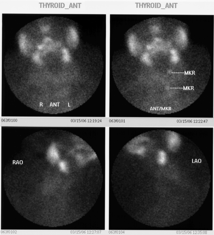
This 74year old female presents with a multinodular goiter. Thyroid scan is shown. Markers were placed on the sternal notch. She was injected with 8.3mci of 99mTc pertechnitate. Images in the A-P (upper panel) and in right anterior oblique (lower panel right) and left anterior oblique (lower panel left). The images show minimal uptake in the by the enlarged gland. There is normal uptake in the salivary glands These findings are consistent with a benign non toxic multinodular thyroid gland.
Courtesy Alan Ashare MD Copyright 2010 95696bb02


    [nodeType] => 1
    [parentNode] => (object value omitted)
    [childNodes] => (object value omitted)
    [firstChild] => (object value omitted)
    [lastChild] => (object value omitted)
    [previousSibling] => (object value omitted)
    [nextSibling] => (object value omitted)
    [attributes] => (object value omitted)
    [ownerDocument] => (object value omitted)
    [namespaceURI] =>
    [prefix] =>
    [localName] => table
    [baseURI] =>
    [textContent] =>


Multinodular Goiter  – Minimal Uptake


This 74year old female presents with a multinodular goiter. Thyroid scan is shown. Markers were placed on the sternal notch. She was injected with 8.3mci of 99mTc pertechnitate. Images in the A-P (upper panel) and in right anterior oblique (lower panel right) and left anterior oblique (lower panel left). The images show minimal uptake in the by the enlarged gland. There is normal uptake in the salivary glands These findings are consistent with a benign non toxic multinodular thyroid gland.
Courtesy Alan Ashare MD Copyright 2010 95696bb02


)
DOMElement Object
(
    [schemaTypeInfo] =>
    [tagName] => td
    [firstElementChild] => (object value omitted)
    [lastElementChild] => (object value omitted)
    [childElementCount] => 2
    [previousElementSibling] =>
    [nextElementSibling] =>
    [nodeName] => td
    [nodeValue] =>
This 74year old female presents with a multinodular goiter. Thyroid scan is shown. Markers were placed on the sternal notch. She was injected with 8.3mci of 99mTc pertechnitate. Images in the A-P (upper panel) and in right anterior oblique (lower panel right) and left anterior oblique (lower panel left). The images show minimal uptake in the by the enlarged gland. There is normal uptake in the salivary glands These findings are consistent with a benign non toxic multinodular thyroid gland.
Courtesy Alan Ashare MD Copyright 2010 95696bb02

    [nodeType] => 1
    [parentNode] => (object value omitted)
    [childNodes] => (object value omitted)
    [firstChild] => (object value omitted)
    [lastChild] => (object value omitted)
    [previousSibling] => (object value omitted)
    [nextSibling] => (object value omitted)
    [attributes] => (object value omitted)
    [ownerDocument] => (object value omitted)
    [namespaceURI] =>
    [prefix] =>
    [localName] => td
    [baseURI] =>
    [textContent] =>
This 74year old female presents with a multinodular goiter. Thyroid scan is shown. Markers were placed on the sternal notch. She was injected with 8.3mci of 99mTc pertechnitate. Images in the A-P (upper panel) and in right anterior oblique (lower panel right) and left anterior oblique (lower panel left). The images show minimal uptake in the by the enlarged gland. There is normal uptake in the salivary glands These findings are consistent with a benign non toxic multinodular thyroid gland.
Courtesy Alan Ashare MD Copyright 2010 95696bb02

)
DOMElement Object
(
    [schemaTypeInfo] =>
    [tagName] => td
    [firstElementChild] => (object value omitted)
    [lastElementChild] => (object value omitted)
    [childElementCount] => 2
    [previousElementSibling] =>
    [nextElementSibling] =>
    [nodeName] => td
    [nodeValue] =>
Multinodular Goiter  – Minimal Uptake
    [nodeType] => 1
    [parentNode] => (object value omitted)
    [childNodes] => (object value omitted)
    [firstChild] => (object value omitted)
    [lastChild] => (object value omitted)
    [previousSibling] => (object value omitted)
    [nextSibling] => (object value omitted)
    [attributes] => (object value omitted)
    [ownerDocument] => (object value omitted)
    [namespaceURI] =>
    [prefix] =>
    [localName] => td
    [baseURI] =>
    [textContent] =>
Multinodular Goiter  – Minimal Uptake
)
DOMElement Object
(
    [schemaTypeInfo] =>
    [tagName] => table
    [firstElementChild] => (object value omitted)
    [lastElementChild] => (object value omitted)
    [childElementCount] => 1
    [previousElementSibling] => (object value omitted)
    [nextElementSibling] => (object value omitted)
    [nodeName] => table
    [nodeValue] =>


Normal Technetium Pertechnitate Scan


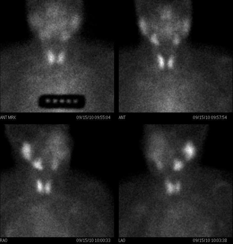
This is a thyroid scan from a 39 year old female with a clinical history of hyperthyroidism. She was injected with 16.1mCi of 99mTechnetium pertechnitate. The scan shows normal uniform uptake of the radioisotope in both lobes of the thyroid gland. The uptake in the thyroid gland is greater than the uptake in the submandibular and parotid glands. These findings are consistent with a normal thyroid gland.
Courtesy Alan Ashare MD Copyright 2010 97511b01


    [nodeType] => 1
    [parentNode] => (object value omitted)
    [childNodes] => (object value omitted)
    [firstChild] => (object value omitted)
    [lastChild] => (object value omitted)
    [previousSibling] => (object value omitted)
    [nextSibling] => (object value omitted)
    [attributes] => (object value omitted)
    [ownerDocument] => (object value omitted)
    [namespaceURI] =>
    [prefix] =>
    [localName] => table
    [baseURI] =>
    [textContent] =>


Normal Technetium Pertechnitate Scan


This is a thyroid scan from a 39 year old female with a clinical history of hyperthyroidism. She was injected with 16.1mCi of 99mTechnetium pertechnitate. The scan shows normal uniform uptake of the radioisotope in both lobes of the thyroid gland. The uptake in the thyroid gland is greater than the uptake in the submandibular and parotid glands. These findings are consistent with a normal thyroid gland.
Courtesy Alan Ashare MD Copyright 2010 97511b01


)
DOMElement Object
(
    [schemaTypeInfo] =>
    [tagName] => td
    [firstElementChild] => (object value omitted)
    [lastElementChild] => (object value omitted)
    [childElementCount] => 2
    [previousElementSibling] =>
    [nextElementSibling] =>
    [nodeName] => td
    [nodeValue] =>
This is a thyroid scan from a 39 year old female with a clinical history of hyperthyroidism. She was injected with 16.1mCi of 99mTechnetium pertechnitate. The scan shows normal uniform uptake of the radioisotope in both lobes of the thyroid gland. The uptake in the thyroid gland is greater than the uptake in the submandibular and parotid glands. These findings are consistent with a normal thyroid gland.
Courtesy Alan Ashare MD Copyright 2010 97511b01

    [nodeType] => 1
    [parentNode] => (object value omitted)
    [childNodes] => (object value omitted)
    [firstChild] => (object value omitted)
    [lastChild] => (object value omitted)
    [previousSibling] => (object value omitted)
    [nextSibling] => (object value omitted)
    [attributes] => (object value omitted)
    [ownerDocument] => (object value omitted)
    [namespaceURI] =>
    [prefix] =>
    [localName] => td
    [baseURI] =>
    [textContent] =>
This is a thyroid scan from a 39 year old female with a clinical history of hyperthyroidism. She was injected with 16.1mCi of 99mTechnetium pertechnitate. The scan shows normal uniform uptake of the radioisotope in both lobes of the thyroid gland. The uptake in the thyroid gland is greater than the uptake in the submandibular and parotid glands. These findings are consistent with a normal thyroid gland.
Courtesy Alan Ashare MD Copyright 2010 97511b01

)
DOMElement Object
(
    [schemaTypeInfo] =>
    [tagName] => td
    [firstElementChild] => (object value omitted)
    [lastElementChild] => (object value omitted)
    [childElementCount] => 2
    [previousElementSibling] =>
    [nextElementSibling] =>
    [nodeName] => td
    [nodeValue] =>
Normal Technetium Pertechnitate Scan
    [nodeType] => 1
    [parentNode] => (object value omitted)
    [childNodes] => (object value omitted)
    [firstChild] => (object value omitted)
    [lastChild] => (object value omitted)
    [previousSibling] => (object value omitted)
    [nextSibling] => (object value omitted)
    [attributes] => (object value omitted)
    [ownerDocument] => (object value omitted)
    [namespaceURI] =>
    [prefix] =>
    [localName] => td
    [baseURI] =>
    [textContent] =>
Normal Technetium Pertechnitate Scan
)

