Intraparietal Sulcus
The Common Vein Copyright 2009
Definition
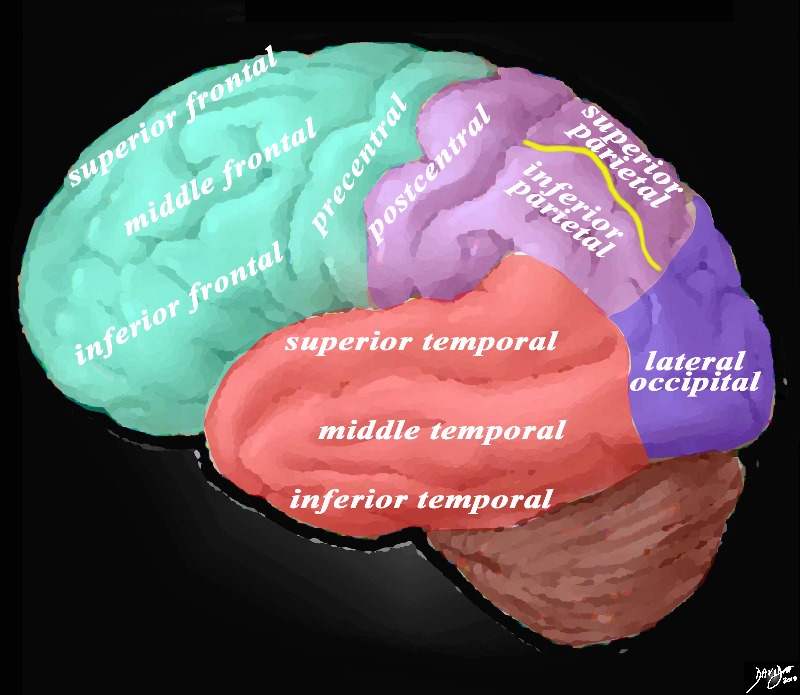
When viewed from the lateral aspect oft the brain three main gyri are seen. The post central gyrus is vertical and lies parallel with the precentral gyrus and central sulcus.
Posterior to the postcentral gyrus are the superior and the inferior parietal lobules.
The intraparietal sulcus runs posteriorly from the postcentral sulcus toward the occipital lobe almost horizontal and it separates the superior and inferior parietal lobules.

Intraparietal Sulcus |
|
The lateral view of the brain shows the frontal lobe (green) parietal lobe (light mauve), the occipital lobe (purple) and the temporal lobe (red) The intraparietal sulcus (yellow) divides the parietal lobe into a superior gyrus and an inferior gyrus. Additionally in this view the frontal lobe gyri that are visible are; superior frontal, middle frontal, inferior frontal and precentral gyri. The parietal gyri include the post central, superior parietal and inferior parietal. The occipital gyrus that is visible is the lateral occipital. The temporal lobe gyri include the superior, middle and inferior temporal gyri.
Courtesy Ashley Davidoff MD Copyright 2010 83029d05g01
|
DOMElement Object
(
    [schemaTypeInfo] =>
    [tagName] => table
    [firstElementChild] => (object value omitted)
    [lastElementChild] => (object value omitted)
    [childElementCount] => 1
    [previousElementSibling] => (object value omitted)
    [nextElementSibling] =>
    [nodeName] => table
    [nodeValue] =>


Intraparietal Sulcus


The lateral view of the brain shows the frontal lobe (green) parietal lobe (light mauve), the occipital lobe (purple) and the temporal lobe (red) The intraparietal sulcus (yellow) divides the parietal lobe into a superior gyrus and an inferior gyrus. Additionally in this view the frontal lobe gyri that are visible are; superior frontal, middle frontal, inferior frontal and precentral gyri. The parietal gyri include the post central, superior parietal and inferior parietal. The occipital gyrus that is visible is the lateral occipital. The temporal lobe gyri include the superior, middle and inferior temporal gyri.
Courtesy Ashley Davidoff MD Copyright 2010 83029d05g01


    [nodeType] => 1
    [parentNode] => (object value omitted)
    [childNodes] => (object value omitted)
    [firstChild] => (object value omitted)
    [lastChild] => (object value omitted)
    [previousSibling] => (object value omitted)
    [nextSibling] => (object value omitted)
    [attributes] => (object value omitted)
    [ownerDocument] => (object value omitted)
    [namespaceURI] =>
    [prefix] =>
    [localName] => table
    [baseURI] =>
    [textContent] =>


Intraparietal Sulcus


The lateral view of the brain shows the frontal lobe (green) parietal lobe (light mauve), the occipital lobe (purple) and the temporal lobe (red) The intraparietal sulcus (yellow) divides the parietal lobe into a superior gyrus and an inferior gyrus. Additionally in this view the frontal lobe gyri that are visible are; superior frontal, middle frontal, inferior frontal and precentral gyri. The parietal gyri include the post central, superior parietal and inferior parietal. The occipital gyrus that is visible is the lateral occipital. The temporal lobe gyri include the superior, middle and inferior temporal gyri.
Courtesy Ashley Davidoff MD Copyright 2010 83029d05g01


)
DOMElement Object
(
    [schemaTypeInfo] =>
    [tagName] => td
    [firstElementChild] => (object value omitted)
    [lastElementChild] => (object value omitted)
    [childElementCount] => 2
    [previousElementSibling] =>
    [nextElementSibling] =>
    [nodeName] => td
    [nodeValue] =>
The lateral view of the brain shows the frontal lobe (green) parietal lobe (light mauve), the occipital lobe (purple) and the temporal lobe (red) The intraparietal sulcus (yellow) divides the parietal lobe into a superior gyrus and an inferior gyrus. Additionally in this view the frontal lobe gyri that are visible are; superior frontal, middle frontal, inferior frontal and precentral gyri. The parietal gyri include the post central, superior parietal and inferior parietal. The occipital gyrus that is visible is the lateral occipital. The temporal lobe gyri include the superior, middle and inferior temporal gyri.
Courtesy Ashley Davidoff MD Copyright 2010 83029d05g01

    [nodeType] => 1
    [parentNode] => (object value omitted)
    [childNodes] => (object value omitted)
    [firstChild] => (object value omitted)
    [lastChild] => (object value omitted)
    [previousSibling] => (object value omitted)
    [nextSibling] => (object value omitted)
    [attributes] => (object value omitted)
    [ownerDocument] => (object value omitted)
    [namespaceURI] =>
    [prefix] =>
    [localName] => td
    [baseURI] =>
    [textContent] =>
The lateral view of the brain shows the frontal lobe (green) parietal lobe (light mauve), the occipital lobe (purple) and the temporal lobe (red) The intraparietal sulcus (yellow) divides the parietal lobe into a superior gyrus and an inferior gyrus. Additionally in this view the frontal lobe gyri that are visible are; superior frontal, middle frontal, inferior frontal and precentral gyri. The parietal gyri include the post central, superior parietal and inferior parietal. The occipital gyrus that is visible is the lateral occipital. The temporal lobe gyri include the superior, middle and inferior temporal gyri.
Courtesy Ashley Davidoff MD Copyright 2010 83029d05g01

)
DOMElement Object
(
    [schemaTypeInfo] =>
    [tagName] => td
    [firstElementChild] => (object value omitted)
    [lastElementChild] => (object value omitted)
    [childElementCount] => 2
    [previousElementSibling] =>
    [nextElementSibling] =>
    [nodeName] => td
    [nodeValue] =>
Intraparietal Sulcus
    [nodeType] => 1
    [parentNode] => (object value omitted)
    [childNodes] => (object value omitted)
    [firstChild] => (object value omitted)
    [lastChild] => (object value omitted)
    [previousSibling] => (object value omitted)
    [nextSibling] => (object value omitted)
    [attributes] => (object value omitted)
    [ownerDocument] => (object value omitted)
    [namespaceURI] =>
    [prefix] =>
    [localName] => td
    [baseURI] =>
    [textContent] =>
Intraparietal Sulcus
)

