The ileocecal valve is the functional valve that separates the small bowel from the large bowel and it is positioned between the cecum and the ascending colon. It is usually surrounded by fat which can sometimes become quite prominent
There are two lips to the valve ? an upper and a lower. Each lip of the valve is formed by a doubling back of the mucosa and circular muscle of the bowel wall, while the longitudinal fibers of the small bowel consolidate into the three bands of taenia coli muscle. Additionally fat accumulates in the submucosa giving the lips of the valve some bulk. The peritoneal covering of the small bowel is also reflected off the small bowel as it continues as a cover of the cecum and small bowel. The mucosal pattern of the small bowel is replaced by the mucosal pattern of the colon as the transition is made.
The valve controls the volume of chyme that enters the large bowel and is both under neural control as well as hormonal control. After ingestion of a meal gastrin, a hormone produced by the stomach, causes the valve to relax and enables the small bowel to empty the chyme into the colon. The valve also prevents reflux of stool content into the small bowel. Mechanical forces caused by the distended cecum serve to reinforce the valve. During a barium study it is not uncommon to visualize the ileocecal valve opening and closing and close a number of times.
 Ileocecal valve Ileocecal valve |
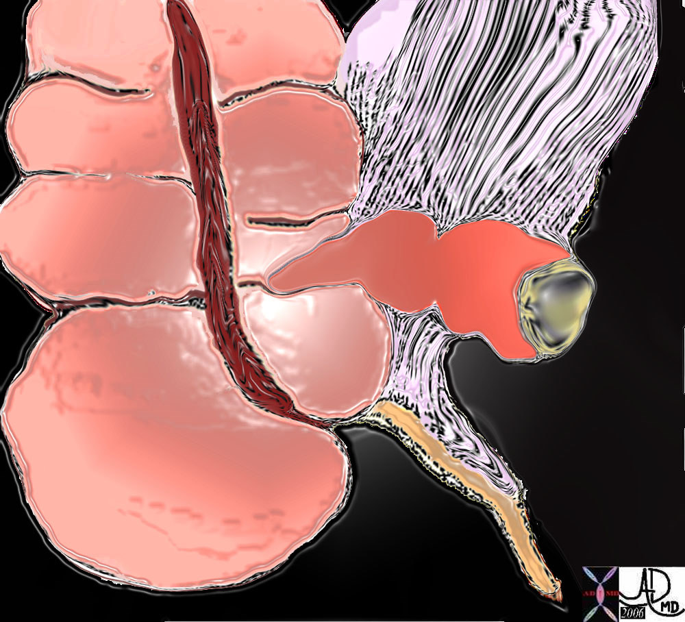
| The diagram shows the junction of the ileum (dark salmon) with the colon.
The ileum marks the border between the cecum inferiorly and the ascending colon superiorly.
The ileocolic ligament (white with black stripes) supports the terminal ileum and is also attached to the ascending colon. The mesoappendix is the mesentery of the appendix and is attached to the vermiform appendix.
Courtesy Ashley Davidoff MD
44657b05 |
 Normal ileocecal valve Normal ileocecal valve |
| This anatomical specimen shows the fatty ileocecal valve (ICV) and the ileum entering the large bowel.
Courtesy Barbara Banner MD
12231 |
 Ileocecal valve Ileocecal valve |
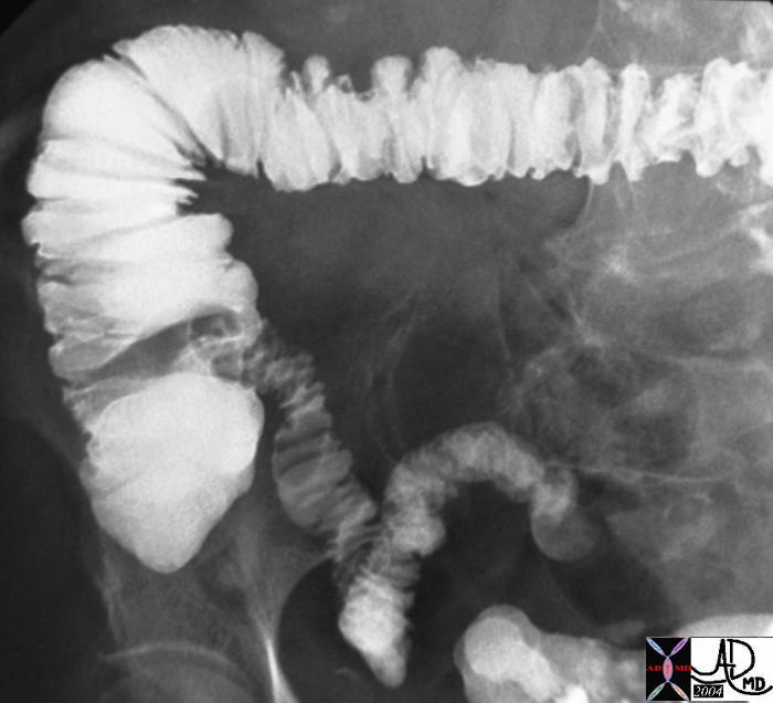
| The end of the road for the small bowel and the beginning of the colon is distinct in this image from a single contrast barium enema. The small bowel has a smaller caliber with terminal ileum in typical location in the right lower quadrant, while the cecum is characterized by a typical conical shape with the cone pointing inferiorly. The ileocecal valve is situated medially, while the ascending colon frames the lateral wall of the right side of the abdomen. In this case the ileocecal valve proved to be incompetent with the barium spilling over. This is not an uncommon finding during the performance of a barium enema.
Courtesy Ashley Davidoff MD
32525 |
The volume of chyme that passes through the ileocecal valve is relatively small and amounts to between 450 and 1000 ccs per day.
 Ileocecal valve and cecum Ileocecal valve and cecum |
| This image reflects the terminal ileum and ileocecal valve in action as demonstrated by a small bowel follow through study. In the first image the valve is open and in the second it is closed. The sphincter enables the valve to close and resist pressures of up to 50-60cms.of water. After a meal the sphincter responds to circulating gastrin by relaxing, and allowing small bowel contents to be emptied into the cecum and thus providing space for the newly ingested meal.
Courtesy Ashley Davidoff MD
26875 |
  Ileocecal valve ? normal CTscan Ileocecal valve ? normal CTscan |
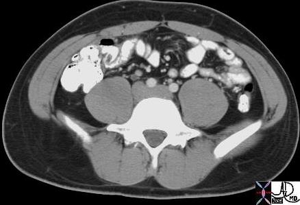
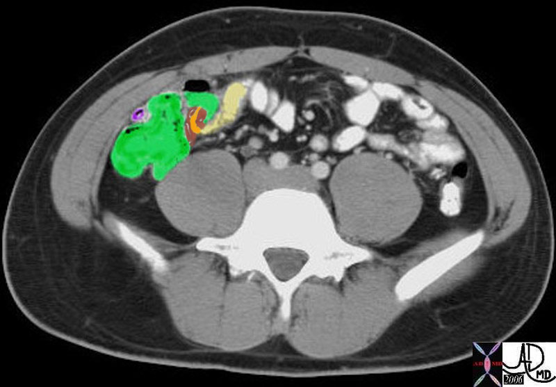
| This CT image shows a normal contrast filled ileum, ileocecal valve (maroon) surrounding the terminal ileum (orange) tucked within the cecum (green) and the extracecal terminal ileum in yellow. The appendix is seen in purple.
Courtesy Ashley Davidoff MD
19491 19491b02 |
Applied anatomy
The most common aberrance of the ileocecal valve is a condition called lipomatous change of the valve where excessive fat is deposited in the valve. This usually has no clinical significance. and is seen by the endoscopist as a submucosal mass. Sometimes Crohn?s disease and ulcerative colitis may affect the valve resulting in a patulous and incompetent valve. Backwash ileitis is an inflammation of the ileum though to be due to an incompetent valve in ulcerative colitis (UC)
 Lipomatous change of the ileocecal valve Lipomatous change of the ileocecal valve |
| The ileocecal valve in this 80 year old female is surrounded by more adipose tissue than usual characterized by a black ring around the terminal ileum. This is not an unusual nor abnormal finding and is called lipomatous change of the ileocecal valve
Courtesy Ashley Davidoff MD
29439 |
DOMElement Object
(
    [schemaTypeInfo] =>
    [tagName] => table
    [firstElementChild] => (object value omitted)
    [lastElementChild] => (object value omitted)
    [childElementCount] => 1
    [previousElementSibling] => (object value omitted)
    [nextElementSibling] =>
    [nodeName] => table
    [nodeValue] =>


Lipomatous change of the ileocecal valve


The ileocecal valve in this 80 year old female is surrounded by more adipose tissue than usual characterized by a black ring around the terminal ileum.  This is not an unusual nor abnormal finding and is called lipomatous change of the ileocecal valve
Courtesy Ashley Davidoff MD
29439


    [nodeType] => 1
    [parentNode] => (object value omitted)
    [childNodes] => (object value omitted)
    [firstChild] => (object value omitted)
    [lastChild] => (object value omitted)
    [previousSibling] => (object value omitted)
    [nextSibling] => (object value omitted)
    [attributes] => (object value omitted)
    [ownerDocument] => (object value omitted)
    [namespaceURI] =>
    [prefix] =>
    [localName] => table
    [baseURI] =>
    [textContent] =>


Lipomatous change of the ileocecal valve


The ileocecal valve in this 80 year old female is surrounded by more adipose tissue than usual characterized by a black ring around the terminal ileum.  This is not an unusual nor abnormal finding and is called lipomatous change of the ileocecal valve
Courtesy Ashley Davidoff MD
29439


)
DOMElement Object
(
    [schemaTypeInfo] =>
    [tagName] => td
    [firstElementChild] => (object value omitted)
    [lastElementChild] => (object value omitted)
    [childElementCount] => 2
    [previousElementSibling] =>
    [nextElementSibling] =>
    [nodeName] => td
    [nodeValue] => The ileocecal valve in this 80 year old female is surrounded by more adipose tissue than usual characterized by a black ring around the terminal ileum.  This is not an unusual nor abnormal finding and is called lipomatous change of the ileocecal valve
Courtesy Ashley Davidoff MD
29439
    [nodeType] => 1
    [parentNode] => (object value omitted)
    [childNodes] => (object value omitted)
    [firstChild] => (object value omitted)
    [lastChild] => (object value omitted)
    [previousSibling] => (object value omitted)
    [nextSibling] => (object value omitted)
    [attributes] => (object value omitted)
    [ownerDocument] => (object value omitted)
    [namespaceURI] =>
    [prefix] =>
    [localName] => td
    [baseURI] =>
    [textContent] => The ileocecal valve in this 80 year old female is surrounded by more adipose tissue than usual characterized by a black ring around the terminal ileum.  This is not an unusual nor abnormal finding and is called lipomatous change of the ileocecal valve
Courtesy Ashley Davidoff MD
29439
)
DOMElement Object
(
    [schemaTypeInfo] =>
    [tagName] => td
    [firstElementChild] => (object value omitted)
    [lastElementChild] => (object value omitted)
    [childElementCount] => 1
    [previousElementSibling] =>
    [nextElementSibling] =>
    [nodeName] => td
    [nodeValue] => Lipomatous change of the ileocecal valve
    [nodeType] => 1
    [parentNode] => (object value omitted)
    [childNodes] => (object value omitted)
    [firstChild] => (object value omitted)
    [lastChild] => (object value omitted)
    [previousSibling] => (object value omitted)
    [nextSibling] => (object value omitted)
    [attributes] => (object value omitted)
    [ownerDocument] => (object value omitted)
    [namespaceURI] =>
    [prefix] =>
    [localName] => td
    [baseURI] =>
    [textContent] => Lipomatous change of the ileocecal valve
)
https://beta.thecommonvein.net/wp-content/uploads/2023/05/29439.jpg
DOMElement Object
(
    [schemaTypeInfo] =>
    [tagName] => table
    [firstElementChild] => (object value omitted)
    [lastElementChild] => (object value omitted)
    [childElementCount] => 1
    [previousElementSibling] => (object value omitted)
    [nextElementSibling] => (object value omitted)
    [nodeName] => table
    [nodeValue] =>


Ileocecal valve ? normal CTscan


This CT image shows a normal contrast filled ileum, ileocecal valve (maroon) surrounding the terminal ileum (orange) tucked within the cecum  (green) and the extracecal terminal ileum in yellow.  The appendix is seen in purple.
Courtesy Ashley Davidoff MD
19491 19491b02


    [nodeType] => 1
    [parentNode] => (object value omitted)
    [childNodes] => (object value omitted)
    [firstChild] => (object value omitted)
    [lastChild] => (object value omitted)
    [previousSibling] => (object value omitted)
    [nextSibling] => (object value omitted)
    [attributes] => (object value omitted)
    [ownerDocument] => (object value omitted)
    [namespaceURI] =>
    [prefix] =>
    [localName] => table
    [baseURI] =>
    [textContent] =>


Ileocecal valve ? normal CTscan


This CT image shows a normal contrast filled ileum, ileocecal valve (maroon) surrounding the terminal ileum (orange) tucked within the cecum  (green) and the extracecal terminal ileum in yellow.  The appendix is seen in purple.
Courtesy Ashley Davidoff MD
19491 19491b02


)
DOMElement Object
(
    [schemaTypeInfo] =>
    [tagName] => td
    [firstElementChild] => (object value omitted)
    [lastElementChild] => (object value omitted)
    [childElementCount] => 2
    [previousElementSibling] =>
    [nextElementSibling] =>
    [nodeName] => td
    [nodeValue] => This CT image shows a normal contrast filled ileum, ileocecal valve (maroon) surrounding the terminal ileum (orange) tucked within the cecum  (green) and the extracecal terminal ileum in yellow.  The appendix is seen in purple.
Courtesy Ashley Davidoff MD
19491 19491b02
    [nodeType] => 1
    [parentNode] => (object value omitted)
    [childNodes] => (object value omitted)
    [firstChild] => (object value omitted)
    [lastChild] => (object value omitted)
    [previousSibling] => (object value omitted)
    [nextSibling] => (object value omitted)
    [attributes] => (object value omitted)
    [ownerDocument] => (object value omitted)
    [namespaceURI] =>
    [prefix] =>
    [localName] => td
    [baseURI] =>
    [textContent] => This CT image shows a normal contrast filled ileum, ileocecal valve (maroon) surrounding the terminal ileum (orange) tucked within the cecum  (green) and the extracecal terminal ileum in yellow.  The appendix is seen in purple.
Courtesy Ashley Davidoff MD
19491 19491b02
)
DOMElement Object
(
    [schemaTypeInfo] =>
    [tagName] => td
    [firstElementChild] => (object value omitted)
    [lastElementChild] => (object value omitted)
    [childElementCount] => 1
    [previousElementSibling] =>
    [nextElementSibling] =>
    [nodeName] => td
    [nodeValue] => Ileocecal valve ? normal CTscan
    [nodeType] => 1
    [parentNode] => (object value omitted)
    [childNodes] => (object value omitted)
    [firstChild] => (object value omitted)
    [lastChild] => (object value omitted)
    [previousSibling] => (object value omitted)
    [nextSibling] => (object value omitted)
    [attributes] => (object value omitted)
    [ownerDocument] => (object value omitted)
    [namespaceURI] =>
    [prefix] =>
    [localName] => td
    [baseURI] =>
    [textContent] => Ileocecal valve ? normal CTscan
)
https://beta.thecommonvein.net/wp-content/uploads/2023/05/19491b02.jpg https://beta.thecommonvein.net/wp-content/uploads/2023/05/19491.jpg
DOMElement Object
(
    [schemaTypeInfo] =>
    [tagName] => table
    [firstElementChild] => (object value omitted)
    [lastElementChild] => (object value omitted)
    [childElementCount] => 1
    [previousElementSibling] => (object value omitted)
    [nextElementSibling] => (object value omitted)
    [nodeName] => table
    [nodeValue] =>


Ileocecal valve and cecum


This image reflects the terminal ileum and ileocecal valve in action as demonstrated by a small bowel follow through study.  In the first image the valve is open and in the second it is closed. The sphincter enables the valve to close and resist pressures of up to 50-60cms.of water.  After a meal the sphincter responds to circulating gastrin by relaxing, and allowing small bowel contents to be emptied into the cecum and thus providing space for the newly ingested meal.
Courtesy Ashley Davidoff MD
26875


    [nodeType] => 1
    [parentNode] => (object value omitted)
    [childNodes] => (object value omitted)
    [firstChild] => (object value omitted)
    [lastChild] => (object value omitted)
    [previousSibling] => (object value omitted)
    [nextSibling] => (object value omitted)
    [attributes] => (object value omitted)
    [ownerDocument] => (object value omitted)
    [namespaceURI] =>
    [prefix] =>
    [localName] => table
    [baseURI] =>
    [textContent] =>


Ileocecal valve and cecum


This image reflects the terminal ileum and ileocecal valve in action as demonstrated by a small bowel follow through study.  In the first image the valve is open and in the second it is closed. The sphincter enables the valve to close and resist pressures of up to 50-60cms.of water.  After a meal the sphincter responds to circulating gastrin by relaxing, and allowing small bowel contents to be emptied into the cecum and thus providing space for the newly ingested meal.
Courtesy Ashley Davidoff MD
26875


)
DOMElement Object
(
    [schemaTypeInfo] =>
    [tagName] => td
    [firstElementChild] => (object value omitted)
    [lastElementChild] => (object value omitted)
    [childElementCount] => 2
    [previousElementSibling] =>
    [nextElementSibling] =>
    [nodeName] => td
    [nodeValue] => This image reflects the terminal ileum and ileocecal valve in action as demonstrated by a small bowel follow through study.  In the first image the valve is open and in the second it is closed. The sphincter enables the valve to close and resist pressures of up to 50-60cms.of water.  After a meal the sphincter responds to circulating gastrin by relaxing, and allowing small bowel contents to be emptied into the cecum and thus providing space for the newly ingested meal.
Courtesy Ashley Davidoff MD
26875
    [nodeType] => 1
    [parentNode] => (object value omitted)
    [childNodes] => (object value omitted)
    [firstChild] => (object value omitted)
    [lastChild] => (object value omitted)
    [previousSibling] => (object value omitted)
    [nextSibling] => (object value omitted)
    [attributes] => (object value omitted)
    [ownerDocument] => (object value omitted)
    [namespaceURI] =>
    [prefix] =>
    [localName] => td
    [baseURI] =>
    [textContent] => This image reflects the terminal ileum and ileocecal valve in action as demonstrated by a small bowel follow through study.  In the first image the valve is open and in the second it is closed. The sphincter enables the valve to close and resist pressures of up to 50-60cms.of water.  After a meal the sphincter responds to circulating gastrin by relaxing, and allowing small bowel contents to be emptied into the cecum and thus providing space for the newly ingested meal.
Courtesy Ashley Davidoff MD
26875
)
DOMElement Object
(
    [schemaTypeInfo] =>
    [tagName] => td
    [firstElementChild] => (object value omitted)
    [lastElementChild] => (object value omitted)
    [childElementCount] => 1
    [previousElementSibling] =>
    [nextElementSibling] =>
    [nodeName] => td
    [nodeValue] => Ileocecal valve and cecum
    [nodeType] => 1
    [parentNode] => (object value omitted)
    [childNodes] => (object value omitted)
    [firstChild] => (object value omitted)
    [lastChild] => (object value omitted)
    [previousSibling] => (object value omitted)
    [nextSibling] => (object value omitted)
    [attributes] => (object value omitted)
    [ownerDocument] => (object value omitted)
    [namespaceURI] =>
    [prefix] =>
    [localName] => td
    [baseURI] =>
    [textContent] => Ileocecal valve and cecum
)
https://beta.thecommonvein.net/wp-content/uploads/2023/05/26875.jpg
DOMElement Object
(
    [schemaTypeInfo] =>
    [tagName] => table
    [firstElementChild] => (object value omitted)
    [lastElementChild] => (object value omitted)
    [childElementCount] => 1
    [previousElementSibling] => (object value omitted)
    [nextElementSibling] => (object value omitted)
    [nodeName] => table
    [nodeValue] =>


Ileocecal valve


The end of the road for the small bowel and the beginning of the colon is distinct in this image from a single contrast barium enema.  The small bowel has a smaller caliber with terminal ileum in typical location in the right lower quadrant, while the cecum is characterized by a typical conical shape with the cone pointing inferiorly. The ileocecal valve is situated medially, while the ascending colon frames the lateral wall of the right side of the abdomen.  In this case the ileocecal valve proved to be incompetent with the barium spilling over.  This is not an uncommon finding during the performance of a barium enema.
Courtesy Ashley Davidoff MD
32525


    [nodeType] => 1
    [parentNode] => (object value omitted)
    [childNodes] => (object value omitted)
    [firstChild] => (object value omitted)
    [lastChild] => (object value omitted)
    [previousSibling] => (object value omitted)
    [nextSibling] => (object value omitted)
    [attributes] => (object value omitted)
    [ownerDocument] => (object value omitted)
    [namespaceURI] =>
    [prefix] =>
    [localName] => table
    [baseURI] =>
    [textContent] =>


Ileocecal valve


The end of the road for the small bowel and the beginning of the colon is distinct in this image from a single contrast barium enema.  The small bowel has a smaller caliber with terminal ileum in typical location in the right lower quadrant, while the cecum is characterized by a typical conical shape with the cone pointing inferiorly. The ileocecal valve is situated medially, while the ascending colon frames the lateral wall of the right side of the abdomen.  In this case the ileocecal valve proved to be incompetent with the barium spilling over.  This is not an uncommon finding during the performance of a barium enema.
Courtesy Ashley Davidoff MD
32525


)
DOMElement Object
(
    [schemaTypeInfo] =>
    [tagName] => td
    [firstElementChild] => (object value omitted)
    [lastElementChild] => (object value omitted)
    [childElementCount] => 2
    [previousElementSibling] =>
    [nextElementSibling] =>
    [nodeName] => td
    [nodeValue] => The end of the road for the small bowel and the beginning of the colon is distinct in this image from a single contrast barium enema.  The small bowel has a smaller caliber with terminal ileum in typical location in the right lower quadrant, while the cecum is characterized by a typical conical shape with the cone pointing inferiorly. The ileocecal valve is situated medially, while the ascending colon frames the lateral wall of the right side of the abdomen.  In this case the ileocecal valve proved to be incompetent with the barium spilling over.  This is not an uncommon finding during the performance of a barium enema.
Courtesy Ashley Davidoff MD
32525
    [nodeType] => 1
    [parentNode] => (object value omitted)
    [childNodes] => (object value omitted)
    [firstChild] => (object value omitted)
    [lastChild] => (object value omitted)
    [previousSibling] => (object value omitted)
    [nextSibling] => (object value omitted)
    [attributes] => (object value omitted)
    [ownerDocument] => (object value omitted)
    [namespaceURI] =>
    [prefix] =>
    [localName] => td
    [baseURI] =>
    [textContent] => The end of the road for the small bowel and the beginning of the colon is distinct in this image from a single contrast barium enema.  The small bowel has a smaller caliber with terminal ileum in typical location in the right lower quadrant, while the cecum is characterized by a typical conical shape with the cone pointing inferiorly. The ileocecal valve is situated medially, while the ascending colon frames the lateral wall of the right side of the abdomen.  In this case the ileocecal valve proved to be incompetent with the barium spilling over.  This is not an uncommon finding during the performance of a barium enema.
Courtesy Ashley Davidoff MD
32525
)
DOMElement Object
(
    [schemaTypeInfo] =>
    [tagName] => td
    [firstElementChild] => (object value omitted)
    [lastElementChild] => (object value omitted)
    [childElementCount] => 1
    [previousElementSibling] =>
    [nextElementSibling] =>
    [nodeName] => td
    [nodeValue] => Ileocecal valve
    [nodeType] => 1
    [parentNode] => (object value omitted)
    [childNodes] => (object value omitted)
    [firstChild] => (object value omitted)
    [lastChild] => (object value omitted)
    [previousSibling] => (object value omitted)
    [nextSibling] => (object value omitted)
    [attributes] => (object value omitted)
    [ownerDocument] => (object value omitted)
    [namespaceURI] =>
    [prefix] =>
    [localName] => td
    [baseURI] =>
    [textContent] => Ileocecal valve
)
https://beta.thecommonvein.net/wp-content/uploads/2023/05/32525.jpg
DOMElement Object
(
    [schemaTypeInfo] =>
    [tagName] => table
    [firstElementChild] => (object value omitted)
    [lastElementChild] => (object value omitted)
    [childElementCount] => 1
    [previousElementSibling] => (object value omitted)
    [nextElementSibling] => (object value omitted)
    [nodeName] => table
    [nodeValue] =>


Normal ileocecal valve


This anatomical specimen shows the fatty ileocecal valve (ICV) and the ileum entering the large bowel.
Courtesy Barbara Banner MD
12231


    [nodeType] => 1
    [parentNode] => (object value omitted)
    [childNodes] => (object value omitted)
    [firstChild] => (object value omitted)
    [lastChild] => (object value omitted)
    [previousSibling] => (object value omitted)
    [nextSibling] => (object value omitted)
    [attributes] => (object value omitted)
    [ownerDocument] => (object value omitted)
    [namespaceURI] =>
    [prefix] =>
    [localName] => table
    [baseURI] =>
    [textContent] =>


Normal ileocecal valve


This anatomical specimen shows the fatty ileocecal valve (ICV) and the ileum entering the large bowel.
Courtesy Barbara Banner MD
12231


)
DOMElement Object
(
    [schemaTypeInfo] =>
    [tagName] => td
    [firstElementChild] => (object value omitted)
    [lastElementChild] => (object value omitted)
    [childElementCount] => 2
    [previousElementSibling] =>
    [nextElementSibling] =>
    [nodeName] => td
    [nodeValue] => This anatomical specimen shows the fatty ileocecal valve (ICV) and the ileum entering the large bowel.
Courtesy Barbara Banner MD
12231
    [nodeType] => 1
    [parentNode] => (object value omitted)
    [childNodes] => (object value omitted)
    [firstChild] => (object value omitted)
    [lastChild] => (object value omitted)
    [previousSibling] => (object value omitted)
    [nextSibling] => (object value omitted)
    [attributes] => (object value omitted)
    [ownerDocument] => (object value omitted)
    [namespaceURI] =>
    [prefix] =>
    [localName] => td
    [baseURI] =>
    [textContent] => This anatomical specimen shows the fatty ileocecal valve (ICV) and the ileum entering the large bowel.
Courtesy Barbara Banner MD
12231
)
DOMElement Object
(
    [schemaTypeInfo] =>
    [tagName] => td
    [firstElementChild] => (object value omitted)
    [lastElementChild] => (object value omitted)
    [childElementCount] => 1
    [previousElementSibling] =>
    [nextElementSibling] =>
    [nodeName] => td
    [nodeValue] => Normal ileocecal valve
    [nodeType] => 1
    [parentNode] => (object value omitted)
    [childNodes] => (object value omitted)
    [firstChild] => (object value omitted)
    [lastChild] => (object value omitted)
    [previousSibling] => (object value omitted)
    [nextSibling] => (object value omitted)
    [attributes] => (object value omitted)
    [ownerDocument] => (object value omitted)
    [namespaceURI] =>
    [prefix] =>
    [localName] => td
    [baseURI] =>
    [textContent] => Normal ileocecal valve
)
https://beta.thecommonvein.net/wp-content/uploads/2023/05/12231.jpg
DOMElement Object
(
    [schemaTypeInfo] =>
    [tagName] => table
    [firstElementChild] => (object value omitted)
    [lastElementChild] => (object value omitted)
    [childElementCount] => 1
    [previousElementSibling] => (object value omitted)
    [nextElementSibling] => (object value omitted)
    [nodeName] => table
    [nodeValue] =>


Ileocecal valve


The diagram shows the junction of the ileum (dark salmon) with the colon.
The ileum marks the border between the cecum inferiorly and the ascending colon superiorly.
The ileocolic ligament (white with black stripes) supports the terminal ileum and is also attached to the ascending colon. The mesoappendix is the mesentery of the appendix and  is attached to the vermiform appendix.
Courtesy Ashley Davidoff MD
44657b05


    [nodeType] => 1
    [parentNode] => (object value omitted)
    [childNodes] => (object value omitted)
    [firstChild] => (object value omitted)
    [lastChild] => (object value omitted)
    [previousSibling] => (object value omitted)
    [nextSibling] => (object value omitted)
    [attributes] => (object value omitted)
    [ownerDocument] => (object value omitted)
    [namespaceURI] =>
    [prefix] =>
    [localName] => table
    [baseURI] =>
    [textContent] =>


Ileocecal valve


The diagram shows the junction of the ileum (dark salmon) with the colon.
The ileum marks the border between the cecum inferiorly and the ascending colon superiorly.
The ileocolic ligament (white with black stripes) supports the terminal ileum and is also attached to the ascending colon. The mesoappendix is the mesentery of the appendix and  is attached to the vermiform appendix.
Courtesy Ashley Davidoff MD
44657b05


)
DOMElement Object
(
    [schemaTypeInfo] =>
    [tagName] => td
    [firstElementChild] => (object value omitted)
    [lastElementChild] => (object value omitted)
    [childElementCount] => 4
    [previousElementSibling] =>
    [nextElementSibling] =>
    [nodeName] => td
    [nodeValue] => The diagram shows the junction of the ileum (dark salmon) with the colon.
The ileum marks the border between the cecum inferiorly and the ascending colon superiorly.
The ileocolic ligament (white with black stripes) supports the terminal ileum and is also attached to the ascending colon. The mesoappendix is the mesentery of the appendix and  is attached to the vermiform appendix.
Courtesy Ashley Davidoff MD
44657b05
    [nodeType] => 1
    [parentNode] => (object value omitted)
    [childNodes] => (object value omitted)
    [firstChild] => (object value omitted)
    [lastChild] => (object value omitted)
    [previousSibling] => (object value omitted)
    [nextSibling] => (object value omitted)
    [attributes] => (object value omitted)
    [ownerDocument] => (object value omitted)
    [namespaceURI] =>
    [prefix] =>
    [localName] => td
    [baseURI] =>
    [textContent] => The diagram shows the junction of the ileum (dark salmon) with the colon.
The ileum marks the border between the cecum inferiorly and the ascending colon superiorly.
The ileocolic ligament (white with black stripes) supports the terminal ileum and is also attached to the ascending colon. The mesoappendix is the mesentery of the appendix and  is attached to the vermiform appendix.
Courtesy Ashley Davidoff MD
44657b05
)
DOMElement Object
(
    [schemaTypeInfo] =>
    [tagName] => td
    [firstElementChild] => (object value omitted)
    [lastElementChild] => (object value omitted)
    [childElementCount] => 1
    [previousElementSibling] =>
    [nextElementSibling] =>
    [nodeName] => td
    [nodeValue] => Ileocecal valve
    [nodeType] => 1
    [parentNode] => (object value omitted)
    [childNodes] => (object value omitted)
    [firstChild] => (object value omitted)
    [lastChild] => (object value omitted)
    [previousSibling] => (object value omitted)
    [nextSibling] => (object value omitted)
    [attributes] => (object value omitted)
    [ownerDocument] => (object value omitted)
    [namespaceURI] =>
    [prefix] =>
    [localName] => td
    [baseURI] =>
    [textContent] => Ileocecal valve
)
https://beta.thecommonvein.net/wp-content/uploads/2023/09/44657b05.jpg
 Normal ileocecal valve


