An accessory fissure is an anatomical variation in the lungs where an extra fissure divides the lung tissue into additional lobes or segments beyond the usual lobar divisions.

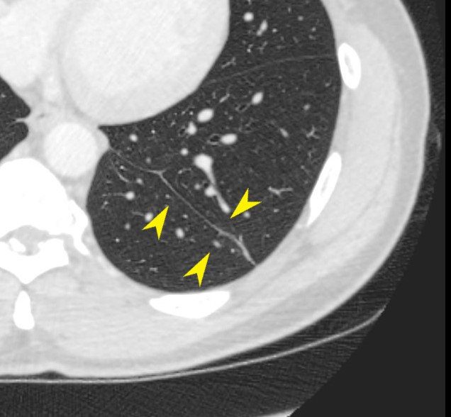
Axial CT scan through the lower chest shows 2 fissures in the left lung. The normal oblique fissure is seen anteriorly and the accessory fissure posteriorly.
Ashley Davidoff TheCommonVein.net
40M-seq

CT scan in axial projection through the lower chest shows the inferior accessory fissure (yellow arrowheads).
Ashley Davidoff TheCommonVein.net
40M-seq
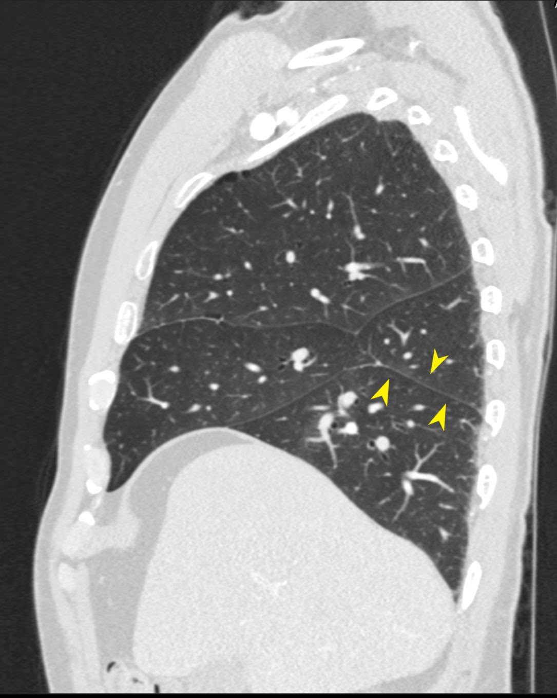
Inferior Accessory Fissure Left Lung

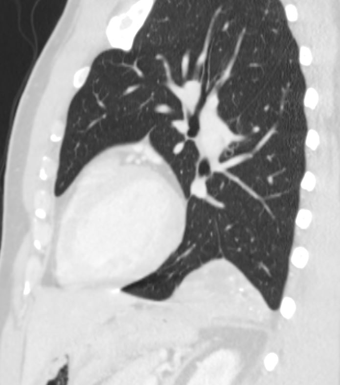
Sagittal CT scan through the left chest shows 2 fissures in the left lung. The normal oblique fissure is seen superiorly and the accessory fissure inferiorly.
Ashley Davidoff TheCommonVein.net
lung-inferior-accessory-fissureL
It is a congenital variation that occurs during lung development. The pathogenesis involves the incomplete fusion of the lung during embryonic development, resulting in an extra cleft of tissue. Accessory fissures do not typically affect lung function but may cause confusion during imaging interpretation, as they can resemble pathological conditions like pleural effusions or scarring.
An accessory fissure refers to an additional anatomical division within the lungs, beyond the standard major fissures. Normally, the lungs are divided into lobes by the following fissures:
- The right lung has two major fissures:
- The oblique fissure, separating the middle and lower lobes.
- The horizontal fissure, separating the upper and middle lobes.
- The left lung has one major fissure:
- The oblique fissure, separating the upper and lower lobes.
An accessory fissure occurs as a developmental variation during embryogenesis, resulting in extra separations within lung tissue. These fissures are visible on imaging studies like chest X-rays or CT scans.
Common types of accessory fissures include:
- Azygos fissure: Found in the right upper lobe and formed by the azygos vein.
- Inferior accessory fissure: Found in the lower lobe, typically separating a segment.
- Superior accessory fissure: Found in the upper lobe, occasionally separating segments.
- Left minor fissure: Analogous to the horizontal fissure but located in the left lung, which is rare.
While accessory fissures are generally benign and have no significant clinical implications, they can influence the interpretation of imaging studies or surgical planning. For instance, they may be mistaken for pathological conditions like pleural effusion or scarring.
(Ref Chat GPT)
Inferior Accessory Fissure

CT scan through the lower chest shows 2 fissures in the left lung. The normal oblique fissure is seen anteriorly and the accessory fissure posteriorly.
Ashley Davidoff TheCommonVein.net
40M-seq

CT scan in axial projection through the lower chest shows the inferior accessory fissure (yellow arrowheads).
Ashley Davidoff TheCommonVein.net
40M-seq

